
Procedure map / Spine
Balloon kyphoplasty
A balloon is inflated inside the vertebral body to create a controlled cavity, which is then filled with bone cement.
Step by step
- 01
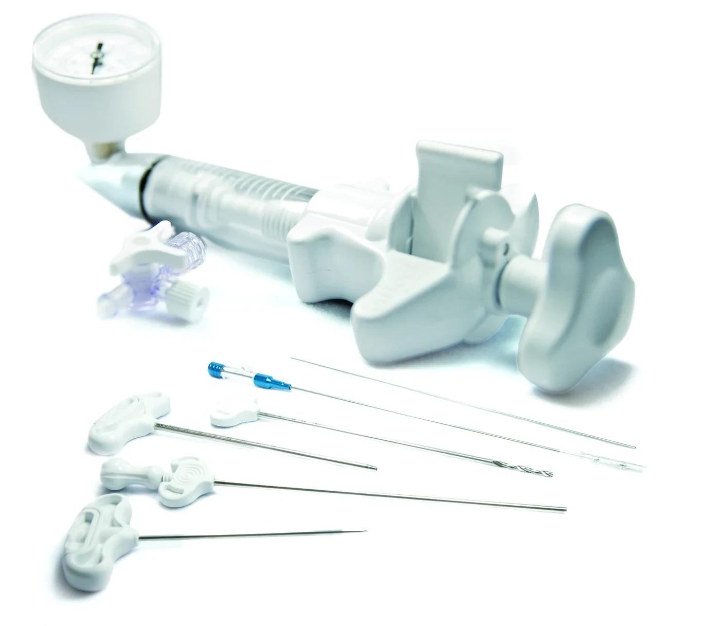
Guide wire and cannula
The bone marrow needle enters the vertebral body through the pedicle and the guide wire is placed; the trocar and cannula are advanced over the wire. Usually done on both sides.
Cannula 178 mm, Ø 4.3 mm
- 02
Opening the balloon path
The bone drill opens the channel for the balloon in the cancellous bone. The channel is then smoothed with the cement filler so sharp bone fragments do not damage the balloon.
Bone drill 220 mm, Ø 3.5 mm
- 03
Balloon inflation
The balloon, filled with contrast, is inflated gradually while pressure and fluoroscopy are watched. Inflation stops once a cavity forms or it reaches the endplates.
Gauge-monitored inflation
- 04
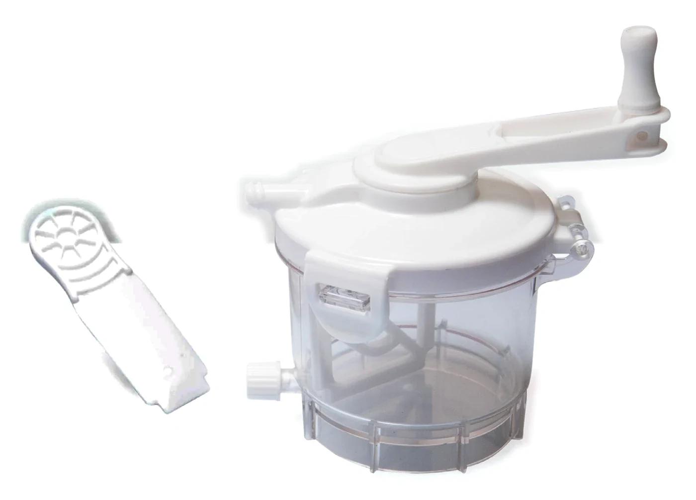
Cement preparation
While the balloon is deflated and withdrawn, the cement is prepared under vacuum. High radiopacity keeps the fill clearly visible on screen.
Radiopacity AI 4.6 mm
- 05
Filling the cavity
The cavity is filled with the cement fillers and pusher under fluoroscopy, then the cannulas are withdrawn.
Cement filler Ø 3-3.5 mm
A general workflow is shown. Practice varies by physician and patient.